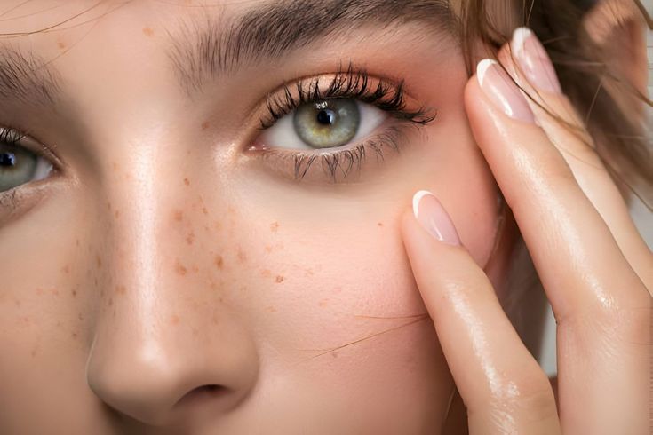
In the realm of dermatology, nestled amidst the intricate fabric of skin conditions, lies a seemingly innocuous yet frequently encountered entity: Milia, colloquially known as Milialar in Turkey. These tiny, pearl-like cysts, though often benign, can pose cosmetic concerns, particularly when they manifest around the delicate periorbital region. Embark on a journey with us as we unravel the enigmatic world of Milia, navigating its multifaceted dimensions, origins, clinical manifestations, and efficacious treatment modalities. Rooted in scientific evidence and clinical insights, our aim is to furnish you with an in-depth comprehension of this dermatological phenomenon, empowering you to make informed decisions regarding your skincare regimen.
Demystifying Milia: An Insightful Overview
What Exactly is Milia or Milialar?
Milia, or Milialar as known in certain regions, manifests as diminutive, dome-shaped bumps typically ranging from 1 to 2 millimeters in size. These cystic formations exhibit a characteristic whitish-yellow hue and possess a firm, smooth consistency upon palpation. Predominantly found in the vicinity of the eyes and eyelids, Milia resemble miniature pearls nestled beneath the skin’s surface. It’s imperative to discern that Milia diverge from acne, as they originate from the entrapment of keratin beneath the skin’s stratum corneum, rather than sebum accumulation within pilosebaceous units.
The Spectrum of Milia: Understanding its Variants
Primary Milia:
Primary Milia, the prototypical form, arises directly from the entrapment of keratin within the skin, predominantly observed in neonates owing to immature sweat ducts. These minute cysts, characterized by their white-to-yellow appearance, often adorn facial regions such as the cheeks, nose, and periorbital area. Remarkably, primary Milia frequently undergo spontaneous resolution within a span of weeks to months, without necessitating therapeutic intervention.
Secondary Milia:
In contrast, secondary Milia ensue subsequent to cutaneous trauma or injury, emerging as a sequelae of dermatological conditions or invasive procedures. Mirroring the characteristics of primary Milia, these cystic lesions are typically localized to the site of insult or intervention. Effective management mandates addressing the underlying etiology, with therapeutic modalities tailored to individual clinical presentations.
Unraveling the Origins: Deciphering Milia Development
Milia genesis ensues when defunct keratinocytes become entrapped beneath the epidermal layer, precipitating the formation of cystic lesions. While they predominantly manifest on facial regions, particularly around the eyes and cheeks, Milia can potentially occur elsewhere on the body. An array of predisposing factors contribute to Milia development, albeit the precise etiological underpinnings may elude elucidation in certain instances. Key contributory factors encompass:
- Genetic Predisposition: Genetic susceptibility plays a pivotal role in Milia pathogenesis, often exhibiting familial aggregation and hereditary transmission of the condition.
- Sun Exposure: Prolonged ultraviolet (UV) radiation exposure exerts deleterious effects on facial integument, augmenting the propensity for Milia formation over time.
- Cutaneous Trauma: Inciting events such as lacerations, burns, or abrasions pave the path for Milia genesis during the reparative phase of wound healing.
- Underlying Dermatological Conditions: Dermatological disorders characterized by xerosis and inflammation, such as eczema, heighten the vulnerability to Milia development.
- Medications: Certain pharmacological agents, notably corticosteroids, may inadvertently precipitate Milia formation as an adverse drug reaction.
- Cosmetic Products: The utilization of occlusive skincare formulations and heavy makeup imparts a propensity for pore occlusion, predisposing to cystic formation and exacerbating extant Milia.
Navigating Therapeutic Avenues: Empowering Milia Management
While Milia often regress spontaneously without therapeutic intervention, persistent or symptomatic cases warrant targeted management strategies. A myriad of treatment modalities exists, tailored to individual preferences and clinical exigencies:
- Topical Retinoid Preparations: Formulations incorporating tretinoin, adapalene, or tazarotene facilitate epidermal desquamation, expediting Milia resolution.
- Microdermabrasion: This minimally invasive procedure employs abrasive crystals to abrade superficial epidermal layers, fostering cellular turnover and mitigating Milia formation.
- Chemical Peels: Utilizing gentle chemical exfoliants such as glycolic or salicylic acid aids in softening and dislodging Milia lesions, fostering a smoother cutaneous texture.
- Electrocautery: Targeted application of electrical current via a hyfrecator cauterizing device enables the precise ablation of Milia lesions, often under local anesthesia.
- Manual Extraction: Dermatological extraction entails incising the cyst with a sterile needle and evacuating its contents, offering immediate relief and aesthetic enhancement.
- Cryotherapy: Cryosurgical techniques involve the controlled application of liquid nitrogen to freeze and eradicate Milia lesions, fostering tissue healing and regeneration.
- Laser Ablation: Laser-assisted Milia removal harnesses focused laser energy to selectively obliterate cystic lesions, yielding precise and cosmetically pleasing outcomes.
- Surgical Excision: In refractory cases, surgical excision may be warranted, involving meticulous removal of Milia lesions under local anesthesia, often necessitating suturing for optimal wound closure.
In Conclusion: Fostering Skin Health and Vitality
Milia, though ostensibly benign, can undermine cutaneous aesthetics and diminish self-esteem. Armed with a comprehensive understanding of Milia pathogenesis, clinical manifestations, and therapeutic modalities, individuals can navigate their skincare journey with confidence and efficacy. By embracing judicious skincare practices, minimizing sun exposure, and seeking timely intervention when warranted, individuals can fortify their defenses against Milia formation and nurture optimal skin health and vitality.
Frequently Asked Questions (FAQs)
Q1. What precisely is Milia?
Milia, characterized by tiny cystic lesions, commonly manifest on the skin, emanating from the entrapment of keratin beneath the epidermal layer. These whitish-yellow protuberances often grace facial regions, particularly around the eyes.
Q2. What triggers the onset of Milia?
Milia may arise from various precipitating factors, including cutaneous trauma, prolonged sun exposure, utilization of occlusive skincare products, certain medications, and genetic predisposition. Neonatal Milia may also ensue in newborns and typically resolve spontaneously.
Q3. How can Milia be preempted?
Preventive measures against Milia entail eschewing heavy or comedogenic skincare formulations that may occlude pores. Incorporating regular exfoliation into skincare routines and shielding the skin from UV damage with sunscreen also play pivotal roles in Milia prevention.
Q4. Are Milia detrimental to health?
In the majority of instances, Milia are benign and devoid of symptomatic manifestations. However, in cases of inflammation or infection, consultation with a dermatologist is advisable for accurate assessment and management.
Q5. What are the treatment options for Milia?
Therapeutic interventions for Milia encompass a spectrum of modalities, including topical retinoid preparations, microdermabrasion, chemical peels, electrocautery, manual extraction, cryotherapy, laser ablation, and surgical excision. The choice of treatment is contingent upon the clinical presentation and individual preferences, with dermatological consultation being instrumental in devising a tailored management plan.

